Surface to Cell: The Real Science of Decomposition
Decomposition is often treated as something to look away from but as a forensic science graduate, I've always found it to be the opposite: a remarkably ordered, well-documented biological sequence. This piece is my attempt to show that, one illustration at a time.
Stage 1 - Fresh
In the hours after death, skin retains its natural colour and texture at first, as the circulation stops a faint paleness sets in. Blood begins to pool in the lowest areas of the body, creating purplish-red skin discoloration, this is an early post-mortem process known as livor mortis.
Without blood flow, cells lose the ability to sustain their own internal environment. Enzymes once safely contained inside the cell's own structures start breaking it down from the inside, this is a process called autolysis.
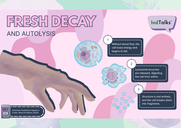
Stage 2 - Bloat
As internal bacteria shift from oxygen-dependent to anaerobic metabolism, they release gases (hydrogen sulphide, methane, and carbon dioxide) that expand abdominal and soft tissues.
The superficial veins stand out as a branching pattern called marbling. As detailed by Cohen, Moss & Prahlow (2025), under the surface, red blood cells have broken down inside the vessel, and their haemoglobin spreads into the surrounding tissue. Bacterial hydrogen sulphide reacting with that haemoglobin is the usual explanation for the colour.
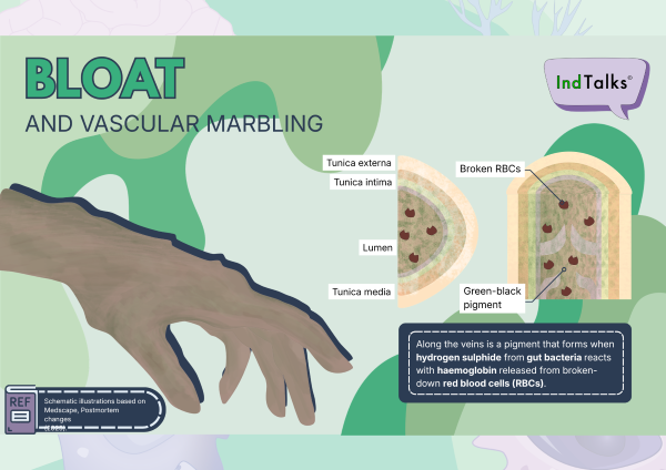
Stage 3 -Active decay
The green of the bloat stage then deepens to dark brown and black. The skin loosens, and the outer layer slips away.
This stage marks a rapid surge in insect activity. As documented by forensic entomology studies (Obafunwa, Roe & Higley 2025), blowflies arrive early to lay eggs. The insect succession is organized into three distinct larval stages (instars), shedding their skin between each. Entomologists read their size and stage to help estimate how long someone has been dead.
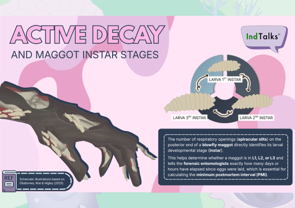
Stage 4 - Advanced decay
Most soft tissue has dissipated, leaving dry, leathery remnants. Decay slows significantly, but the biological focus shifts beneath the body into the soil.
Research into human decomposition (Cobaugh et al., 2015; Carter, Yellowlees & Tibbett, 2007) highlights the creation of a Cadaver Decomposition Island (CDI). Nutrient-rich fluids laden with carbon, nitrogen, and phosphorus saturate the earth. The soil exhibits increased electrical conductivity, altered pH levels, and lower oxygen levels.
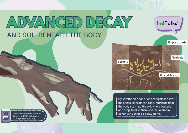
Stage 5 - Dry Remains
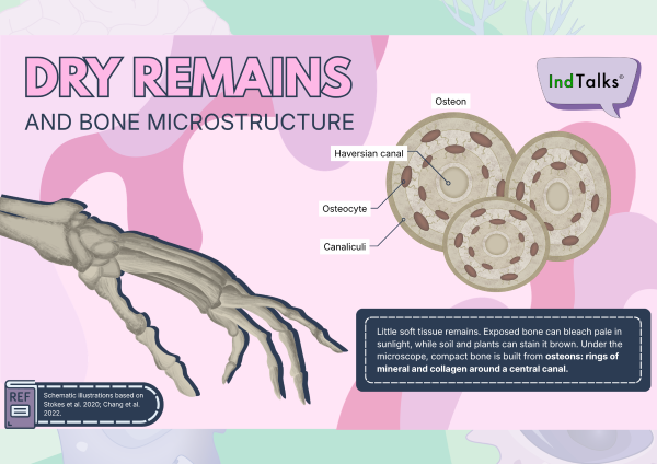
With soft tissues gone, organic matter gives way to durable mineral structures. What remains is bone: bleached white by the sun or stained brownish-tan by soil minerals.
Zooming into the micro-architecture reveals the highly ordered structure of cortical bone. Concentric rings of mineralized matrix (lamellae) form structural units called osteons. At the centre lies the Haversian canal, surrounded by tiny pockets (lacunae) where osteocytes once resided, interconnected by delicate channels (canaliculi). It serves as a quiet, structural testament to the body's enduring chemistry.
Closing
Every stage in this set describes a process, but none of it happens to an abstract body. Each one belongs to a person. That is why the science matters. Understanding how the body changes after death helps investigators estimate how long someone has been gone, which can help to identify them. For the people left behind, identification can mean closure: a name, an answer, a chance to say goodbye.
Forensic science is often told through the “how”. The “why” is the careful, patient work that treats a person with dignity at every stage.
The illustrations in this post are schematic summaries based on the cited sources, not depictions of a single specimen. Timings vary with temperature and conditions.
References
Carter, D.O., Yellowlees, D. & Tibbett, M. (2007). Cadaver decomposition in terrestrial ecosystems. Naturwissenschaften, 94(1), 12-24. https://doi.org/10.1007/s00114-006-0159-1
Cobaugh, K.L., Schaeffer, S.M. & DeBruyn, J.M. (2015). Functional and structural succession of soil microbial communities below decomposing human cadavers. PLoS ONE, 10(6), e0130201. https://doi.org/10.1371/journal.pone.0130201
Cohen PR, Moss RJ Jr, Prahlow JA. Livor mortis and forensic dermatology: a review of death-related gravity-dependent lividity and postmortem hypostasis. Cureus. 2025;17(8):e90760. doi:10.7759/cureus.90760.
Medscape. Postmortem changes. https://emedicine.medscape.com/article/1680032-overview.
Obafunwa J, Roe A, Higley L. A review of the estimation of postmortem interval using forensic entomology. Med Sci Law. 2025;65(1):52-64. doi:10.1177/00258024241275893.
Stokes, S., Márquez-Grant, N. & Greenwood, C.E. (2020). Establishing a minimum PMI for bone sun bleaching in a UK environment with a controlled desert-simulated comparison. International Journal of Legal Medicine, 134(6). https://doi.org/10.1007/s00414-020-02385-y
Comments
Loading comments…